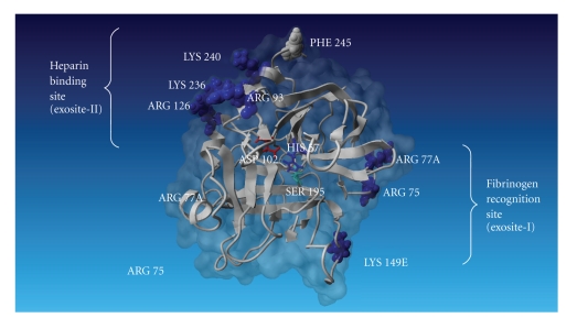
Figure 2.
A model of human thrombin (1 ppb) is represented by ribbon in gray, and molecular surface is represented in dark gray. The catalytic triad composed by His-57, Asp-102, and Ser-195 is shown in the middle of the figure. Heparin-binding site and fibrinogen recognition site are shown at the left and right sides of thrombin, respectively (this image was done by YASARA, reference proteins 47,393-402).

