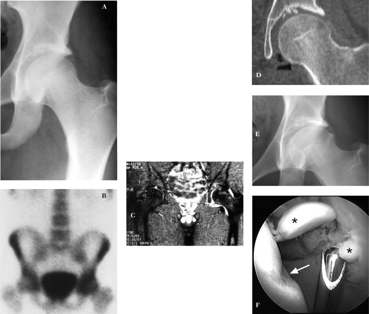
Figure 3.
A. AP radiograph left hip unremarkable. B. Radionuclide scan reveals increased activity, left hip. C. MRI remarkable for pronounced asymmetric effusion, left hip. D. CT coronal reconstruction demonstrates loose bodies. E. Follow-up radiograph 13 months post-injury reveals secondary changes wth superolateral osteophyte formation on the femoral head. F. Loose bodies are evident (*) originating from the acetabulum. Scoring of the femoral head is also evident (arrow) due to third body wear. (Reprinted with permission J.W. Thomas Byrd, M.D.)

