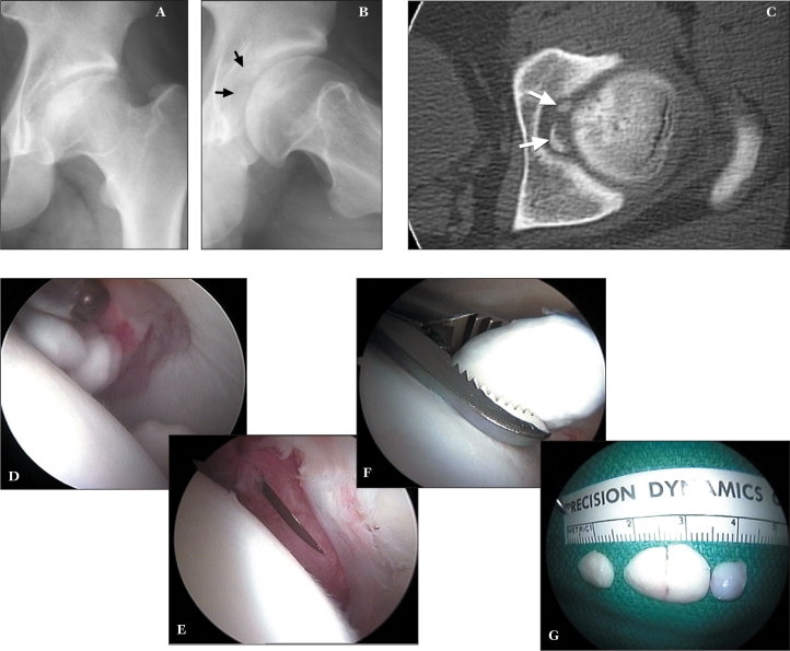
Figure 6.
A 20 year old male with a three month history of acute left hip pain. A. AP radiograph demonstrates findings consistent with old Legg-Calvé Perthes disease. B. Lateral view defines the presence of intraarticular loose bodies (arrows). C. CT scan substantiates the intraarticular location of the fragments (arrows). D. Arthroscopic view medially demonstrates the loose bodies. E. Viewing anteriorly, the anterior capsular incision is enlarged with an arthroscopic knife to facilitate removal of the fragments. F. One of the fragments is being retrieved. G. Loose bodies are able to be removed whole. (Reprinted with permission.3)

