
Figure 2. Subcellular localization of AtVMA21a.
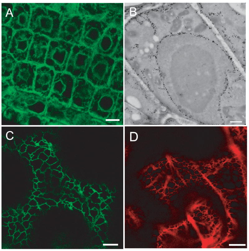
A) AtVMA21a-GFP localizes to the ER in Arabidopsis. CLSM image of root tip cells stably expressing AtVMA21a-GFP under the control of the endogenous promotor, scale bars represent 10μm. B) Overview of an immunogold labeled cryosection of a root tip cortex cell expressing AtVMA21-GFP. Gold markers accumulate in nuclear envelope, ER strands and the cortical ER, scale bar represents 1μm. C) AtVMA21a-GFP localizes to the ER in N. benthamiana. CLSM image of leave epidermal cells transiently expressing AtVMA21a-GFP after Agrobacterium infiltration. D) mRFP-AtVMA21 is also localized to the ER. CLSM image of leave epidermal cells transiently expressing mRFP-AtVMA21a after Agrobacterium infiltration, scale bar represents 10μm.