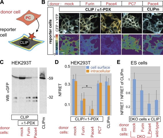
Figure 2.
CLIP detects paracrine PC activity. (A) Strategy to detect cell nonautonomous PC activities. (B) Fluorescent images and heat maps of NFRET in HEK293T cells exposed to donor cells expressing the indicated constructs. Bar, 10 µm. (C) Anti-GFP immunoblotting of conditioned medium from the co-cultures such as those shown in B. (D) Cell nonautonomous PC activity quantified by FRET analysis of CLIP at the surface (blue bars) or in the cytoplasm (orange bars) of the reporter cells shown in B. (E) FRET efficiency of CLIP in Furin−/−;Pace4−/− ES cells (DKO) co-cultured with wild-type ES cells or with separately transfected DKO cells expressing Furin, Pace4, or empty vector. The asterisk indicates a significant difference as determined by t test (P < 0.05). Error bars are means ± SD.

