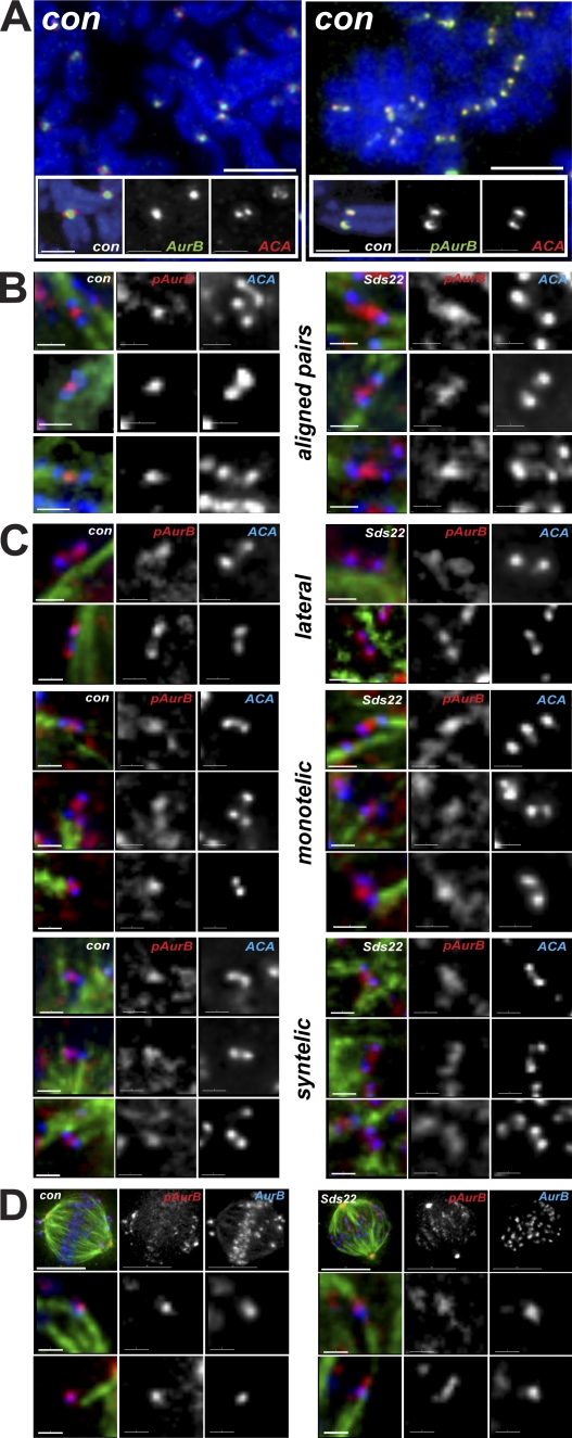
Figure 4.
Localization of phospho–T232 aurora B is determined by Sds22. (A) Localization of aurora B and phospho-T232 aurora B in chromosome spreads after arrest with nocodazole. Phospho-T232 aurora B is concentrated at kinetochores, whereas bulk aurora B remains concentrated at centromeres. Overlay shows DAPI (blue), aurora B or anti–phospho-T232 aurora B (green), and ACA (red). (B) Localization of phospho-T232 aurora B in bioriented chromosomes in fixed intact cells. Sds22 depletion causes phospho-T232 aurora B to spread toward kinetochores. Overlay shows ACA (blue), tubulin (green), and anti–phospho-T232 aurora B (red). (C) Localization of phospho-T232 aurora B in laterally and monooriented chromosomes in fixed intact cells. Phospho-T232 aurora B appears on kinetochores in control and Sds22-depleted cells. Overlay shows ACA (blue), tubulin (green), and anti–phospho-T232 aurora B (red). (D) Colocalization of phospho-T232 aurora B and total aurora B in fixed intact cells. Sds22 depletion causes phospho-T232 aurora B to spread toward kinetochores. Overlay shows phospho-T232 aurora B (red), aurora B (blue), and tubulin (green). Bars: (A, top) 5 µm; (A, insets) 2 µm; (B, C, and D [bottom]) 1 µm; (D, top) 10 µm.

