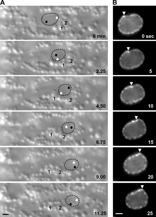
Figure 4.
Actively migrating nuclei roll. (A) DIC images from a time-lapse sequence of nuclear rolling. Nuclear migration is to the right, and rolling is clockwise. The nucleus is outlined; black and white dots represent the position of nucleoli within the nucleus. Two cytoplasmic vesicles (1 and 2) are marked. (B) Images of lamin::GFP from a time-lapse sequence. Rolling is led by a deformation in the lamina (arrowheads). Nuclear migration is to the left, and rolling is clockwise. Bar, 1 µm.

