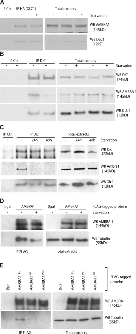
Figure 2.
Modulation of AMBRA1–dynein interaction during autophagy. (A) AMBRA1–DLC1 interaction upon autophagy induction. 2F cells coinfected with retroviral vectors encoding HA-tagged DLC1 and AMBRA1 proteins were nutrient starved for 4 h (+) or left untreated (−). Protein extracts were subjected to IP with an anti-HA–tagged antibody (IP HA; DLC1) or, as a negative control, with an unrelated antibody (IP control [Ctr]). Purified complexes were analyzed together with the corresponding total extracts by WB using anti-AMBRA1 (WB AMBRA1; top) and anti-DLC1 antibodies (WB DLC1; bottom). (B) AMBRA1 dissociates from the dynein motor complex during autophagy. 2F cells were nutrient starved for 4 h or left untreated. Protein extracts were subjected to IP using an anti-DIC (IP DIC) or an unrelated antibody (IP Ctr) as a negative control. The purified complexes were analyzed by WB together with the corresponding total extracts by using anti-DIC (WB DIC; top), anti-AMBRA1 (WB AMBRA1; middle), and anti-DLC1 antibodies (WB DLC1; bottom). (C) AMBRA1 dissociates from the dynein motor complex in mouse tissues after autophagy induction. Mice from the same litter were kept without food for 24 and 48 h or fed ad libitum (−) before sacrifice and necropsy. Kidneys from these mice were homogenized and subjected to IP analysis by using an anti-DIC antibody (IP DIC) or with an unrelated antibody (IP Ctr). Protein immunocomplexes were probed together with the corresponding total extracts using anti-DIC (WB DIC, top), anti-AMBRA1 (WB AMBRA1; middle), and anti-DLC1 (WB DLC1; bottom) antibodies. (D) AMBRA1 dissociates from tubulin upon autophagy induction. 2F cells infected with retroviral vectors encoding Flag-tagged βgal or AMBRA1 proteins were nutrient starved for 4 h or left untreated. Protein extracts were subjected to IP with an anti-Flag antibody (IP Flag). Purified proteins were eluted using the Flag peptide and analyzed by WB together with the corresponding total extracts using anti-AMBRA1 (WB AMBRA1; top) and anti–α-tubulin antibodies (WB tubulin; bottom). (E) AMBRA1 interacts with tubulin via DLC1. 2F cells were coinfected with retroviral vectors encoding Flag-tagged AMBRA1 FL, AMBRA1TAT1, or AMBRA1TAT2. Protein extracts were subjected to IP using an anti-Flag antibody (IP Flag). Purified complexes and corresponding total extracts were analyzed by WB using an anti-AMBRA1 (WB AMBRA1; Top) or anti–α-tubulin antibody (WB tubulin; bottom).

