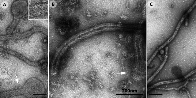
Figure 8.
The incubation of Gth with liposomes at low pH induces the formation of tubular structures. (A) At pH 6, Gth, inserted into liposomes that were initially spherical, forms a local network (indicated by the arrow and enlarged in the top right frame), favoring the formation of tubular structures. (B) At pH 5.5, Gth forms more extensive, regular arrays at the surface of liposomes, resembling those formed by G at the surface of the virus. Spherical vesicles are nevertheless still visible (arrow). (C) At pH 5.2, only rigid tubular protein–lipid structures are observed, at the surface of which Gth displays quasi-helical symmetry. All the samples were negatively stained for electron microscopy observation.

