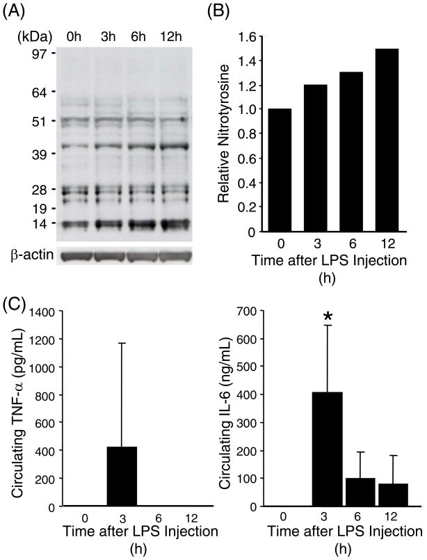
Fig. 1. Increased tyrosine nitration in the lung proteins during systemic inflammation.
(A) Immuno-reactive nitrotyrosine was detected by Western blot analyses on whole lung protein samples from C57BL/6 mice (2-mo-old, male) that were sacrificed at indicated time points after LPS injection (20mg/kg, i.p.). Each lane represents pooled protein samples from 3–4 mice. Immuno-reaction with an anti-β-actin antibody confirms equal loading of the protein samples. (B) Densitometric analysis of (A). Total intensity throughout each lane was normalized to β-actin level. The normalized intensity value of the control lane (0 hour) was set at 1. (C) Plasma concentrations of TNFα and IL-6 were determined in each mouse used for (A). * indicates p<0.05 as compared to 0 hour samples.

