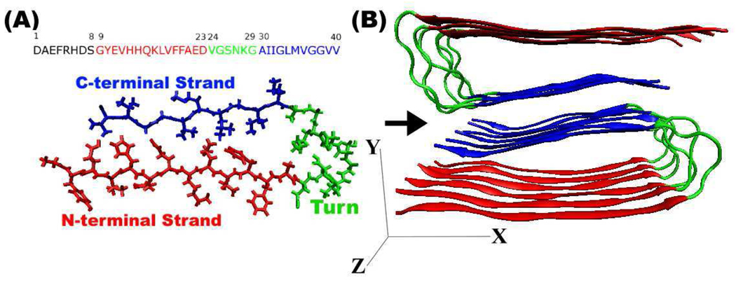
Figure 1.
(A) Aβ1–40 sequence and the structure of a single Aβ9–40 peptide, with asymmetric “U” structure. N-terminal strand (red), C-terminal strand (blue), and turn region (green), respectively. (B) Model structure of amyloid fibril made of 12 Aβ9–40 molecules. Red and blue indicate two parallel β-sheets.

