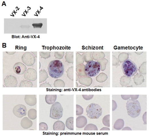
Figure 6. Spatiotemporal Localization of VX-4 by immunocytochemical staining.
(A) rVX-2, rVX-3 and rVX-4 proteins were separated by 12% SDS-PAGE and transferred to PVDF membrane. The membrane was incubated with anti-rVX-4 (1∶1000 dilutions) for 4 h, with an additional incubation with peroxidase-conjugated anti-mouse IgG (1∶2000 dilutions) for 4 h. The blot was developed using 4C1N chromogen. (B) A thin blood smear from a patient with vivax malaria was stained with anti-rVX-4 conjugated with avidin-biotin complex system. The protein was densely labeled in the food vacuoles and adjacent areas, as compared to the staining of the dark hemozoin pigment.

