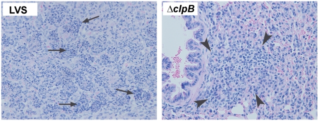
Figure 7. Histopathology of pulmonary tularemia in mice immunized with ΔclpB or LVS.
left: The lung from an LVS-immunized mouse killed at day 7 post challenge showing severe subacute bronchopneumonia. The alveolar spaces are filled with large numbers of intact and degenerated neutrophils (arrows) admixed with small numbers of mononuclear cells. (right) the lung from a clpB-immunized mouse killed at day 7 post challenge showing the infiltration of predominantly mononuclear cells (arrowheads) with virtually no neutrophils in the peribronchial area. H&E, x400.

