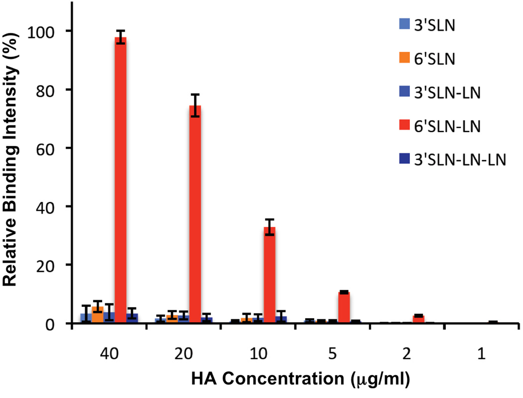
Figure 1. Dose dependent direct receptor binding of CA/04 HA.
A streptavidin plate array comprising representative biotinylated α2–3 and α2–6 sialylated glycans were used for the assay. The biotinylated glycans include 3’SLN, 6’SLN, 3’SLN-LN, 6’SLN-LN and 3’SLN-LN-LN. LN corresponds to lactosamine (Galβ1–4GlcNAc) and 3’SLN and 6’SLN respectively correspond to Neu5Ac α2–3 and Neu5Ac α2–6 linked to LN. The assay was carried out as described previously (18) for an entire range of HA concentration from 0.01 – 40 µg/ml by pre-complexing HA: primary antibody: secondary antibody in the ratio 4:2:1 to enhance the multivalent presentation of HA. The binding signals for HA concentrations below 1 µg/ml were at background level and hence there concentrations are not shown on the x-axis. The y-axis shows the normalized binding signal as a percentage of the maximum value.

