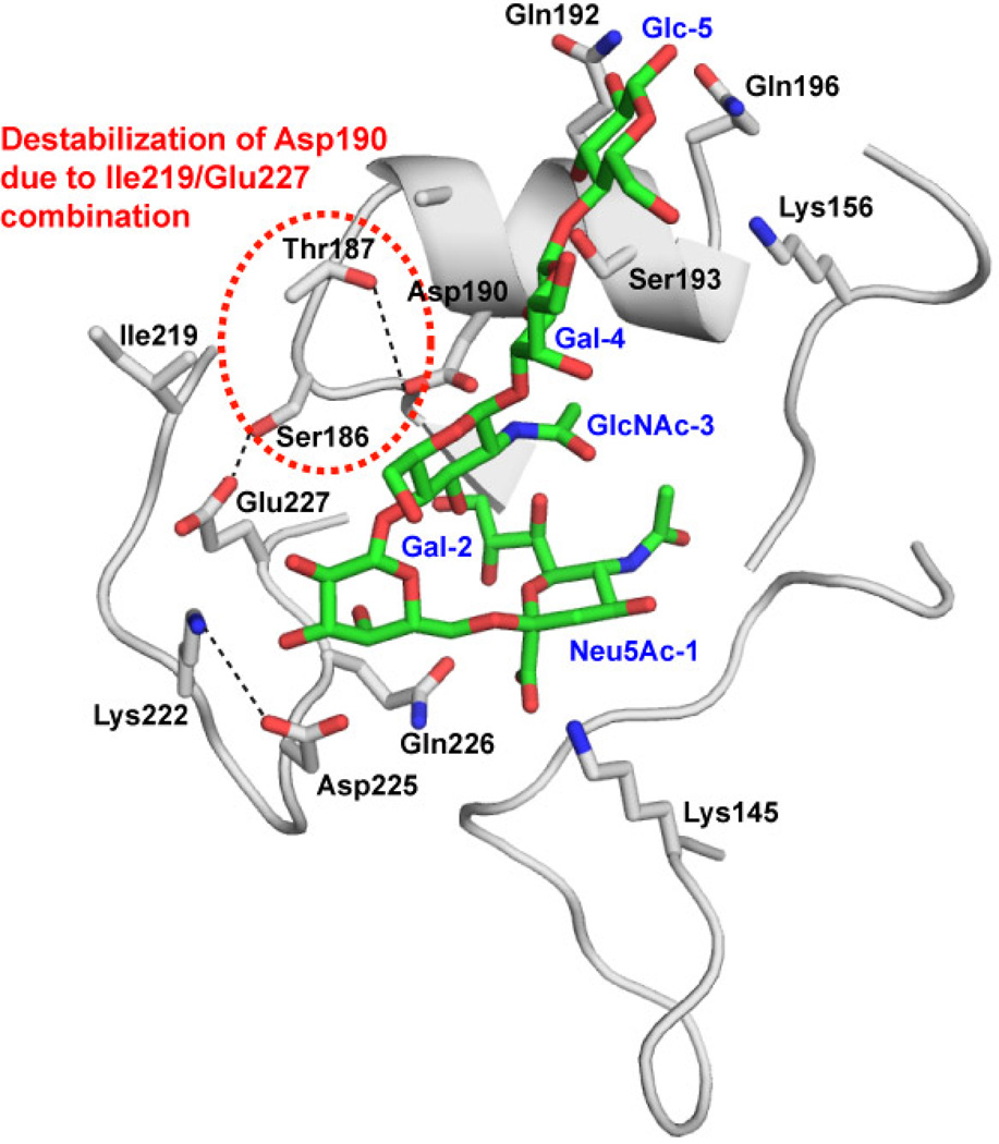
Figure 3. Structural model of CA/04 HA bound to α2–6 oligosaccharide.
The contacts of CA/04 HA with an α2–6 oligosaccharide (Neu5Ac α2–6Galβ1–4GlcNAcβ1–3Galβ1–4Glc) were analyzed by constructing a structural model as described previously (20). Shown in the figure is the cartoon representation of the glycan binding site of CA/04 HA where the side chains of the key amino acids are shown in stick representation (colored by atom carbon: gray; oxygen: red; nitrogen:blue). The α2–6 oligosaccharide is shown as a stick representation (colored by atom carbon: green; oxygen: red; nitrogen:blue) and labeled blue starting from non-reducing end Neu5Ac-1 to reducing end Glc-5. The potential destabilization of the interaction network due to the Ile219/Glu227 combination is highlighted in red dotted circle.

