Figure 1.
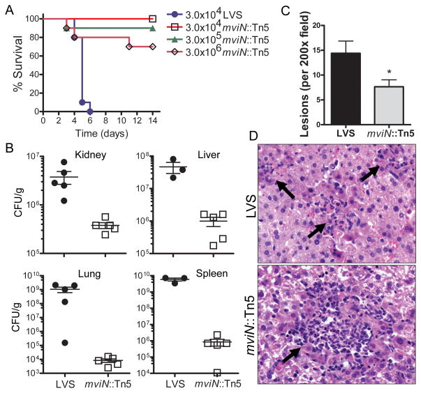
F. tularensis LVS mviN mutant is attenuated in vivo. (A) WT (n=10 per group) mice were injected i.p. with the indicated amount of F. tularensis LVS (LVS) or the mviN mutant (mviN::Tn5) and survival monitored. Data are representative of 5 independent experiments at the 3 × 10⁴ infective dose, each involving a minimum of 5 mice per group. (B) 3 d p.i. with 3 × 10⁴ CFU of LVS or the mviN mutant organs were harvested, homogenized and dilutions plated for enumeration of CFU (n=3–5 mice per group). (C) Number of lesions per 200 × field of H&E-stained liver sections from WT mice 3 d p.i. with either LVS or the mviN mutant were scored; n=5 mice per group (3 random fields were examined per mouse); *, p = 0.016. (D) Representative H&E-stained sections of liver from WT mice 3 d p.i. with either F. tularensis LVS or the mviN mutant. Arrows indicate necropurulent foci.

