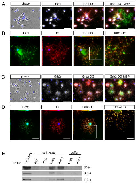
Fig 5. Dystroglycan is found in a protein complex with Grb2 and IRS1.
Oligodendrocytes were differentiated for 3 days on laminin followed by Immunocytochemistry. (A) Immunocytochemistry to visualize IRS1 (white) in conjunction with dystroglycan (red). Co-expression of the mature oligodendrocyte protein, MBP (green), was used to confirm oligodendrocyte identity. Scale bar, 50 μM. (B) Immunocytochemistry to visualize IRS1 (green) in conjunction with dystroglycan (red). Scale bar, 25 μM; 10 μM in inset, right. (C) Immunocytochemistry to visualize Grb2 (white) in conjunction with dystroglycan (red). Co-expression of the mature oligodendrocyte protein, MBP (green), was used to confirm oligodendrocyte identity. Scale bar, 50 μM. (D) Immunocytochemistry to visualize Grb2 (green) in conjunction with dystroglycan (red). Scale bar, 25 μM; 10 μM in inset, right. (E) Immunoprecipitations were performed with lysates obtained from oligodendrocytes differentiated for 3 days (input), using antibodies against Grb2, IRS1, or control rabbit IgG, in conjunction with Protein A/G beads. Immunopreciptation complexes isolated with Grb-2 antibodies contained Grb-2, β-dystroglycan (βDG), and IRS1. Immunopreciptation complexes isolated with IRS-1 antibodies contained IRS1, β-dystroglycan and Grb-2. Samples devoid of antibody (“none”), or treated with the equivalent amount of control antibody (“IgG”) did not isolate detectable levels of Grb-2, β-dystroglycan, or IRS1. Control samples containing equivalent amounts of antibody (Grb-2 or IRS1) but devoid of lysate were included to demonstrate that observed bands were not due to cross-reactivity of Western Blot secondary antibodies with IP antibodies.

