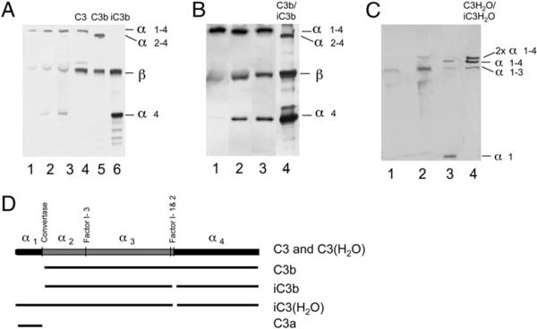
FIGURE 6.
Platelet-associated C3, as detected by Western blot analysis using polyclonal anti-C3c Abs (A, B) or anti-C3a mAb 7SD17.3 (C). A, Platelets were activated by TRAP in PRP for 0, 15, or 30 min. Lanes 1–3 show membrane-bound C3 at the three time points, and lanes 4–6 show control C3, C3b, and iC3b, respectively, at 3 μg each. B, Binding and cleavage of purified C3 after incubation with washed, activated platelets in the absence (lane 1) or presence of factor I (lane 2) or with both factor I and factor H (lane 3). Lane 4 shows a reference comprising a mixture of C3b and iC3b. C, Membrane-bound C3 from nonactivated (lane 1) or TRAP-activated platelets (lane 2). Lane 3 shows control zymosan-activated serum (containing C3a in addition to nonactivated C3), and lane 4 shows control C3(H2O) incubated with factor I and factor H producing iC3(H2O). D, Diagram of the structure of C3 and its various proteolytic activation fragments.

