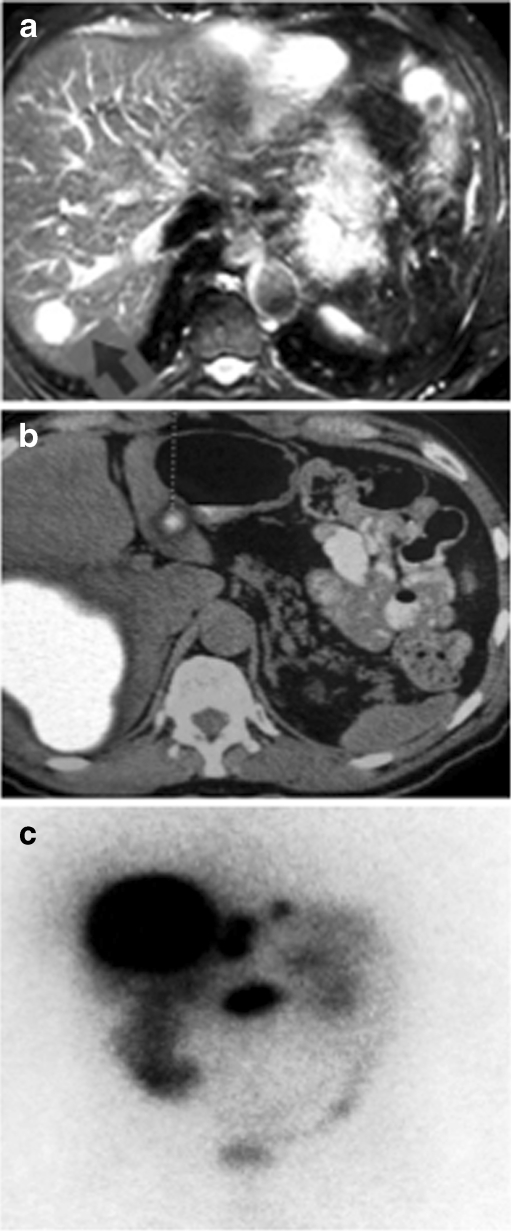
Fig. 1.
a MRI liver in 2004 showing one of the lesions which was hyperintense with a typical ‘light-bulb’ appearance on plain T2-weighted images. b FDG-PET scan showing uptake in the liver and pancreatic lesions with regional lymph node involvement. c OctreoScan confirming metastatic pancreatic neuroendocrine tumor

