
Fig. 6.
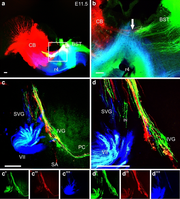
The fiber growth to the ear in E11.5-day embryos. The injection of three different dyes into the cerebellum (CB), red in (a,b), the brainstem green in (a,b) and into rhombomere 4 (r4) efferents, blue in (a,b) reveals a non-overlapping projection to the ear (c,d). Cerebellar afferents to the posterior canal (red in c,d) and brainstem afferents to the posterior canal (green in c,d) are each singly labeled with either color. Likewise, efferents are discreetly labeled (blue in c,d). Consistent with the peripheral labeling, central projections show only few green brainstem fibers to extend beyond the VIIIth nerve root (arrow in b) and only an occasional red cerebellar fiber projects into the brainstem. Combined, these data suggest that the initial fiber growth from sensory neurons to the brain is biased into a population that projects primarily to the brainstem and a population that projects primarily to the cerebellum. Scale bars 100 µm