FIGURE 6.
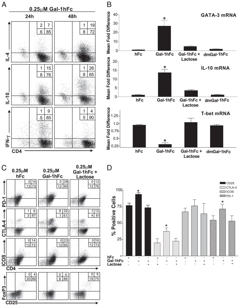
Gal-1hFc alters Th cell differentiation, cytokine production, and expression of regulatory surface molecules. A, Naive mouse Th cells activated with anti-CD3/28 were treated with 0.25 μM Gal-1hFc; and IL-4, IL-10, and IFN-γ for 24 or 48 h were assessed by intracellular cytokine FACS staining. B, Transcriptional activity of GATA-3, IL-10, and T-bet mRNA was analyzed 8 h after incubation with Gal-1hFc by quantitative RT-PCR. Data are expressed as relative mRNA levels normalized to hFc treatment. *Statistically significant difference compared with hFc control, p ≤ 0.01. C, Naive mouse Th cells activated with anti-CD3/28 were incubated for 24 h with 0.25 μM hFc or Gal-1hFc (±50 mM lactose), or stained with anti–CTLA-4, –PD-1, -ICOS, and -CD25 mAbs, and analyzed by flow cytometry. D, Graphic representation of data from three independent experiments. *Statistically significant difference compared with hFc control, p ≤ 0.01.

