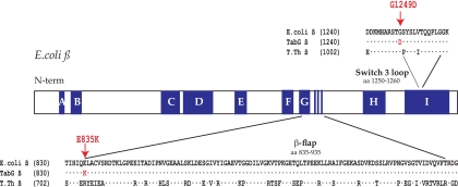
FIG. 1.
Location of TabG rpoB mutations. Schematic representation of the E. coli rpoB gene showing conserved regions (A, B, C, etc.) in dark blue and positions of the TabG substitutions E835K and G1249D within the β flap and adjacent to the switch 3 loop, respectively. Sequences of the residues surrounding the β flap and the switch 3 loop are given for the wt E. coli, for the E. coli TabG and B11, and for the T. thermophilus (T. th.) β proteins.

