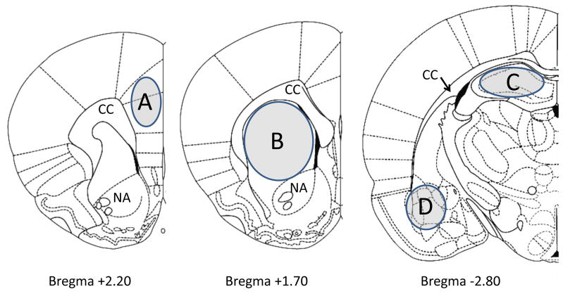
Figure 4.
Schematic representation of coronal sections of the rat brain depicting the regions that were microdissected for analysis via HPLC-ED. The sections were taken from Paxinos and Watson (1998). Shaded portions denote microdissected areas of the (A) prefrontal cortex, (B) striatum, (C) hippocampus, and (D) amygdala. Relevant anatomical structures are: CC, corpus callosum; NA, nucleus accumbens.

