
Figure 2. Elastin, fibrillin-1 and collagen composition of skin biopsies.

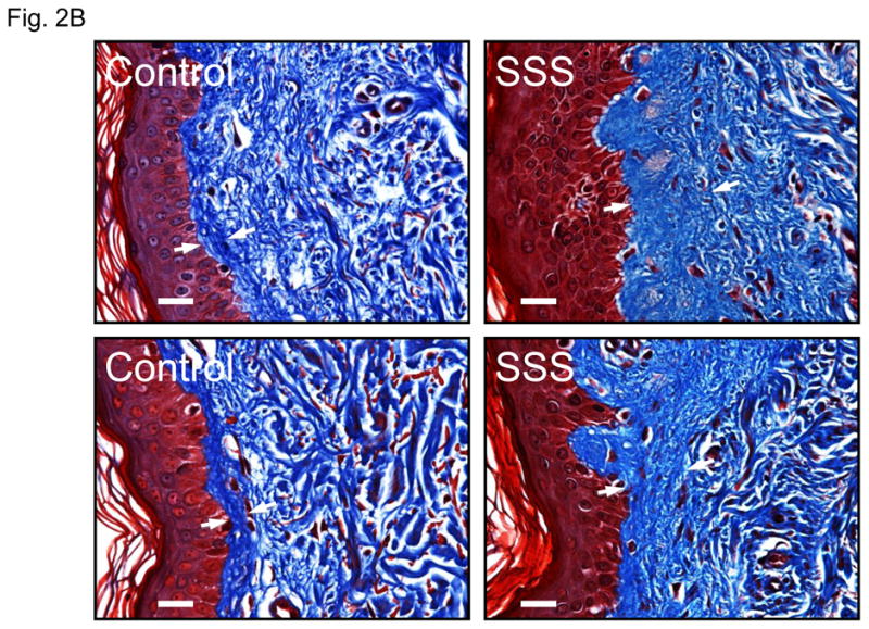
(A) Confocal microscopy of skin biopsies (with keratin in the epithelium delineated in red) from a patient with SSS and a control, seen at low magnification (upper panel) and high magnification (lower panel). The control sample shows long microfibrillar projections from the dermal-epidermal junction (DEJ, arrow) into the superficial (papillary) dermis with relatively sparse fibrillin-1 deposition in the deeper dermis. There is relative exclusion of elastin in the superficial dermis immediately adjacent to the DEJ. The SSS sample shows stubby microfibrillar deposits immediately adjacent to the DEJ that co-localize with elastin. There is also increased deposition of elastin in the deeper dermis. Scale bars = upper row of low magnification images, 50 microns; lower row of high magnification images, 20 microns. (B) Trichrome staining of skin biopsies from two controls and two SSS patients. Note the widened zone of increased deposition of collagen (blue) in the papillary dermis of SSS patients, as delineated by white arrows. Scale bars, 20 microns