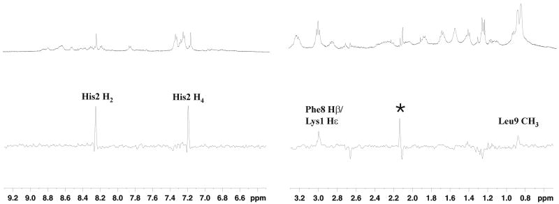
Figure 1.
STD NMR spectrum of Shepherdin peptide in the presence of Hsp90-NT. Upper panel: 1H NMR reference spectrum of 1.2 mM Shepherdin in the presence of 50 μM Hsp90-NT in 30 mM buffer phosphate (95% H2O, 5% D2O), 100 mM NaCl, 6 mM DTT, pH 6.7 recorded at 280K on a 500 MHz Bruker spectrometer. Lower panel: 1H NMR STD spectrum of the same sample. A saturation time of 2 sec at -602 Hz was used. The assignment of the observed STD signals is reported, an asterisk marks a signal due to an impurity (see Material and Methods).

