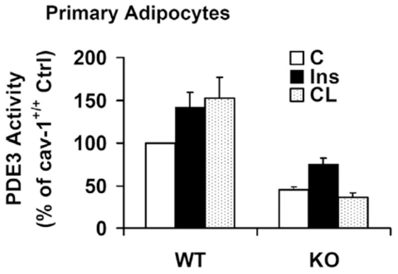
Figure 7. Effects of insulin or CL on PDE3 activity in adipocytes from WT (wild-type) and Cav-1−/− mice.

Adipocytes from Cav-1+/+ (WT) and Cav-1−/− (KO) mice were prepared, incubated without (Ctrl) or with insulin (Ins) (2 nM for 10 min) or CL (10 μM for 15 min), and membrane-associated PDE3 activity was measured as described in the Materials and methods section. Results are presented as the percentage of wild-type control PDE3 activity (means + S.E.M., n = 4–7); absolute values were 20–90 pmol/min per mg of protein.
