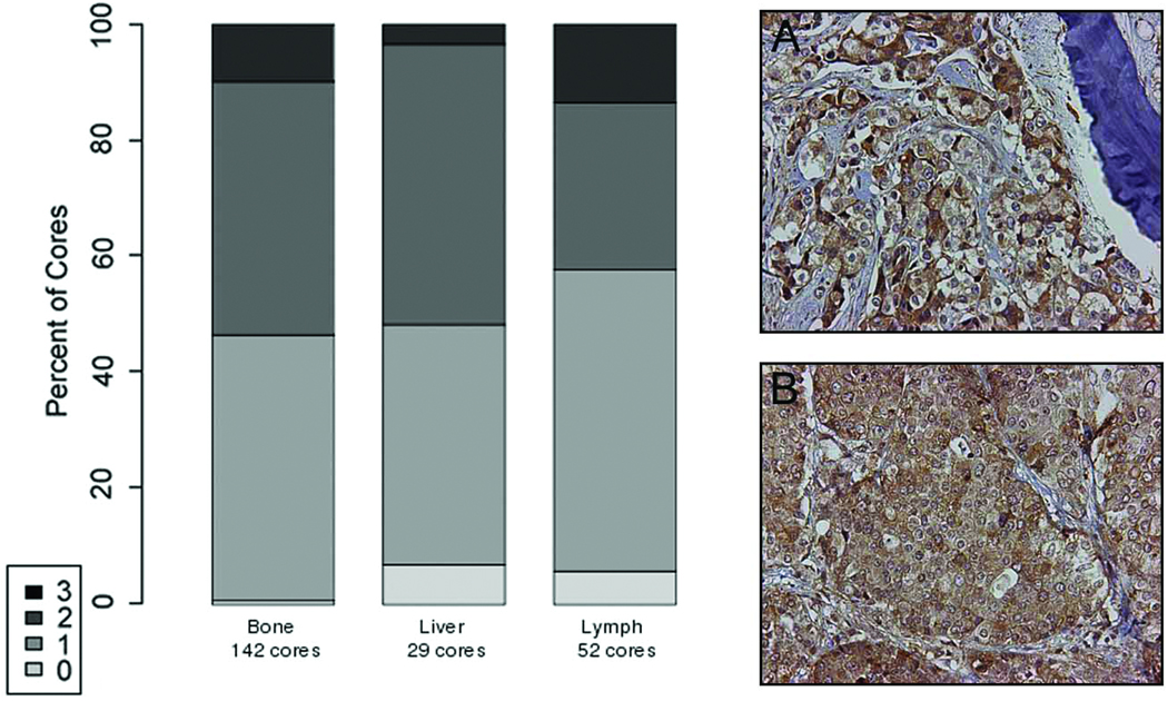
Figure 2. Immunohistochemical analysis of transferrin receptor (TfR) expression.
in human PCa bone (Panel A) and lymph node (Panel B) metastases from 22 patients (200-fold magnification). There was no evidence that TfR staining intensity varies between bone and soft tissue metastases. Specific immunostaining was assessed on a four-point scale: 3 = intense, 2 = defined, 1 = faint, and 0 = absent.

