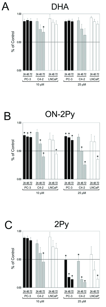
Figure 3. Cell number as assessed by crystal violet assay in LNCaP, C4-2 and PC-3 cells.
Cells were brought to 50% confluence and then cultured in RPMI 1640 with 10% FBS medium with DMSO, 10, or 25 µM DHA, ON-2Py or 2Py for 24, 48, and 72 h. Relative cell number was assessed by crystal violet assay (n = 4). Results are expressed as the mean ± SD. * indicates significant difference from control (p < 0.05).

