
Figure 4. 3 T MRI of a plaque in the right common carotid artery that demonstrates fibrous cap rupture with ulcer formation (yellow arrow).
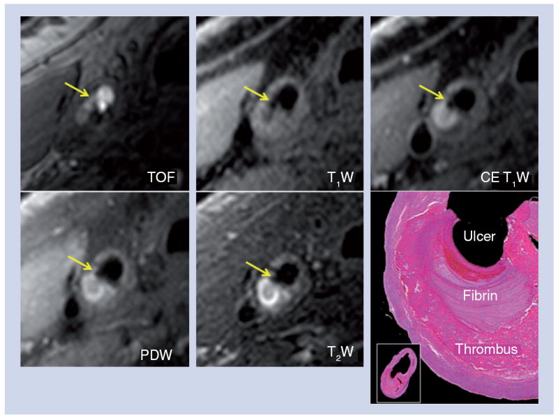
The crescent-shaped, high-signal region in the PDW, T2W and CE T1W images corresponds to a region of thrombus formation, shown on the matched histology section (hematoxylin and eosin stain).
CE: Contrast enhanced; PDW: Proton density weighted; T1W: T1-weighted; T2W: T2-weighted; TOF: Time-of-flight.
Reproduced with permission from [63].