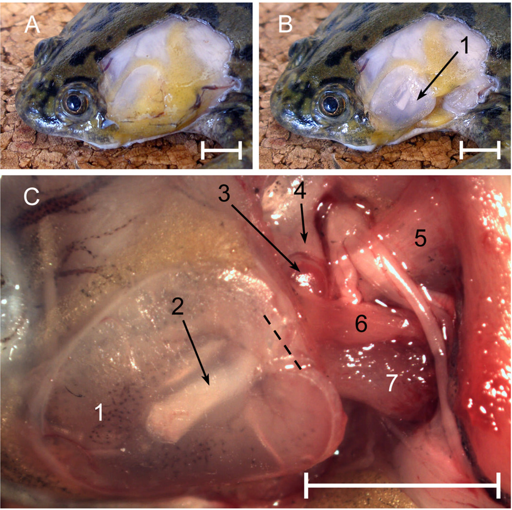
Figure 1.
Left middle ear structures in male Xenopus laevis. A: Skin removed to reveal fatty pad. B: Tympanic disk, after removal of some of the fat. C: Ear region after further dissection, dorsolateral view. Key: 1, tympanic disk; 2, pars media of stapes, seen through disk cartilage; 3, pars interna of stapes; 4, parotic crest; 5, m. levator scapulae superior; 6, m. petrohyoideus; 7, m. cucullaris. Dashed line = approximate position of the rotatory axis. Scale bars represent 5 mm.

