
Figure 1.
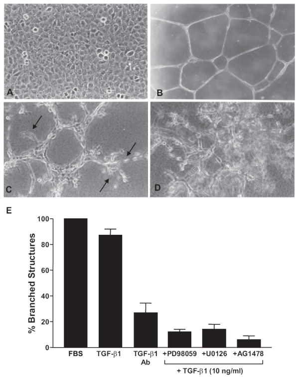
Tubular differentiation of T2 cells in Matrigel culture requires TGF-β1 signaling. T2 cells cultured on dried Matrigel surfaces established contact-inhibited monolayers (A) contrasting with the development of smooth branched tubular networks when seeded onto thin hydrated Matrigel coatings. (B) Cells cultured on thick, hydrated, Matrigel underlays constructed tubular aggregates with extensive sprouting (arrows) evident at terminal foci.(C) Sprout formation and network complexity was most highly developed in 3-D suspension culture in a combination Virtogen:Matrigel support scaffold. (D) Addition of TGF-β1 alone yielded an almost equivalent tubular differentiation response on thin Matrigel matrices compared to FBS supplementation. (E) Incubation with pan-TGF-β antibodies capable of neutralizing TGF-β1, β2, β3 and β5 largely inhibited this response indicating that network formation in the Matrigel system was largely TGF-β-dependent. Pharmacologic blockade of MEK/ERK and EGFR activity with PD98059/U0126 or AG1478, respectively, effectively inhibited TGF-β1-initiated tubular differentiation. Data plotted (in E) is the mean ± SD of triplicate experiments using a calibrated ocular grid to quantify the number of network branchpoints (e.g. B) on thin Matrigel coatings.