
Figure 2.
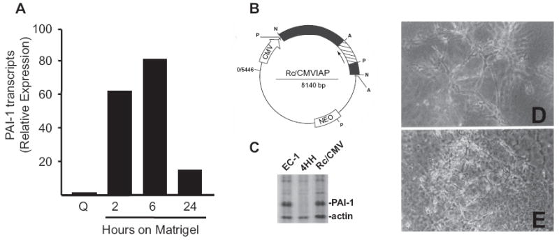
Targeted disruption of PAI-1 expression inhibits formation of stable tubular networks in 3-D Vitrogen:Matrigel suspension culture. PAI-1 transcripts are rapidly up-regulated within 2 hours after plating T2 cells onto thin, hydrated, Matrigel coatings and are maximal at 6 hours corresponding with rapid migration into primitive tube precursor structures. (A) Tubular networks are well-developed by 24 hours at which time PAI-1 mRNA levels remain 18-fold elevated over quiescent (Q) T2 monolayer cultures. Transfection of the Rc/CMVIAP PAI-1 antisense vector (B) into EC-1 cells resulted in derivation of the stable, PAI-1 functionally-null, 4HH cell line. Gel electrophoresis of the 35S-methionine-labeled, matrix-enriched, protein fraction of EC-1, 4HH and empty vector (Rc/CMV)-transfectants confirmed that 4HH cells were, in fact, PAI-1-deficient. (C) Unlike parental and T2 cells that formed stable complex tubular networks in 3-D culture (D), 4HH cells failed to construct stable differentiated structures and effectively degraded the supporting matrix scaffold (E).