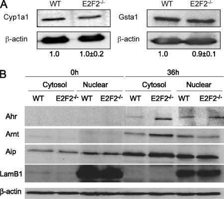
Fig. 5.
(A) Western blot analysis of Cyp1a1 and Gsta1 expression demonstrating comparable protein levels in WT and E2F2−/− T lymphocytes activated through their T-cell receptor. Expression values were normalized against β-actin. The ratios Cyp1a1/β-actin and Gsta1/β-actin in WT were considered as a unit. (B) Western blot analyzes of quiescent (0h) or proliferating (36h) T lymphocytes showing differential subcellular localization of Ahr pathway proteins upon E2F2 loss. Accumulation of Ahr pathway proteins was observed in the cytosolic fraction of E2F2 deficient T lymphocytes. LamB1 was used as nuclear marker and β-actin as loading control. The experiment shown is representative of two independent experiments (n = 6 WT; n = 6 E2F2−/−).

