Fig. 6.
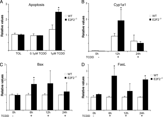
TCDD-mediated activation of Ahr pathway in proliferating T lymphocytes leads to an increased apoptosis in E2F2−/− T cells. T cells were activated using anti-CD3 antibody for 36 hours and subsequently treated with TCDD for the referred times. (A) Apoptosis ratio in the presence of TCDD and vehicle toluene (TOL) of activated WT and E2F2−/− T lymphocytes. (B–D) RT-PCR analysis of Cyp1a1, Bax and FasL in TCDD treated activated T cells derived from WT and E2F2−/− mice (B) Quantitative RT-PCR analysis of Cyp1a1 mRNA expression. Values were normalized against the mRNA expression levels of an irrelevant gene (eEf1a1) and given as relative values. (C, D) Quantitative RT-PCR analysis of the proapoptotic genes FasL and Bax. Values were normalized against the mRNA expression levels of an irrelevant gene (eEf1a1), and relative values obtained in wild-type samples were considered as a unit in each experimental time point (n = 9 WT; n = 9 E2F2−/−; three independent experiments).

