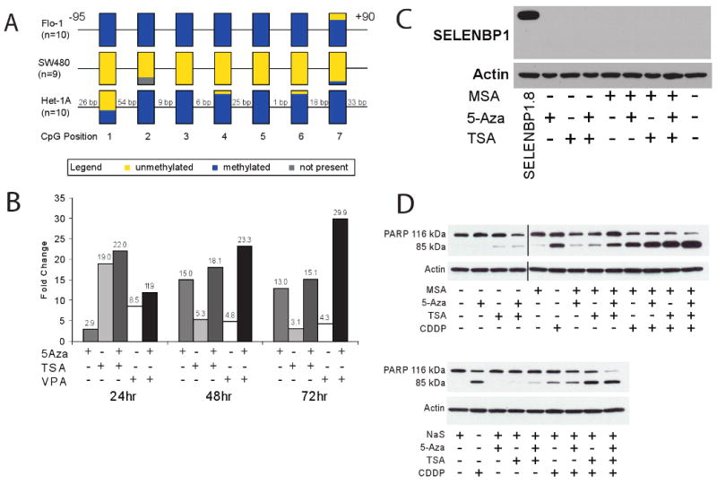
Figure 3.

A, Bisulfite sequencing of Flo-1 (EAC), SW480 (colon adenocarcinoma) and Het-1A (immortalized esophageal squamous epithelium) suggested that hypermethylation of the 5′ upstream promoter region of SELENBP1 occurs in esophageal tissues. B, Treatment of Flo-1 cells with 5 μM 5-aza-2-deoxycytidine (5-Aza), 300 nM trichostatin A (TSA) and 5 mM valproic acid (VPA) induced SELENBP1 expression compared to vehicle-treated controls, as determined by qRT-PCR. C, Treatment of Flo-1 cells with 2.5 μM methylseleninic acid (MSA), 5 μM 5-Aza, and/or 300 nM TSA did not lead to detectable levels of SELENBP1 protein. Stably-transfected SELENBP1.8 cells were used as a positive control. β-actin was used as a loading control. D, Treatment of Flo-1 cells with combinations of 2.5 μM MSA, 5 μM 5-Aza, 300 nM TSA, 10 μM sodium selenite (NaS) and/or 20 μg/mL cisplatin (CDDP) induced apoptosis, as determined by PARP cleavage.
