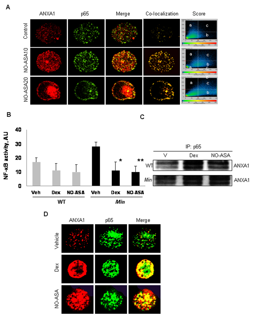
Fig. 5. ANXA1 co-localizes with the p65 subunit of NF-κB in response to NO-ASA treatment.
A: BxPC-3 cells were treated for 2 h with NO-ASA 10 µM (NO-ASA10) or 20 µM (NO-ASA20), fixed with 4% paraformaldehyde and reacted with mouse anti-ANXA1 mAb and anti-p65 rabbit mAb, which do not cross-react with each other, followed by Alexa 555 (anti-mouse; red fluorescence)- and Alexa 488 (anti-rabbit; green fluorescence)-conjugated secondary antibodies and examined by confocal microscopy. Co-localization of the two proteins generates yellow fluorescence. Score graph panels show the fluorescence intensity of p65 alone (a); ANXA1 alone (b); or of both when co-localized (c). B: Min mice (n= 6) and C57BL/6J wild type (WT) mice (n=6) were treated with vehicle (Veh), or NO-ASA 100 mg/kg or dexamethasone 10 mg/kg intraperitoneally, 26 and 2 h before sacrifice. NF-κB activity was determined by ELISA in protein extracts from small intestinal epithelial cells of these mice. Values are mean±SD (*P<0.05 and **P<0.01). C: Protein cell lysates from small intestinal epithelial cells from each animal were immunoprecipitated using an anti-p65 mAb and immunoblotted using an anti-ANXA1 mAb. Representative examples from wild type and Min mice are shown. D: Single small intestinal epithelial cells from Min mice were analyzed by confocal microscopy. Yellow color indicates co-localization of ANXA1 and p65.

