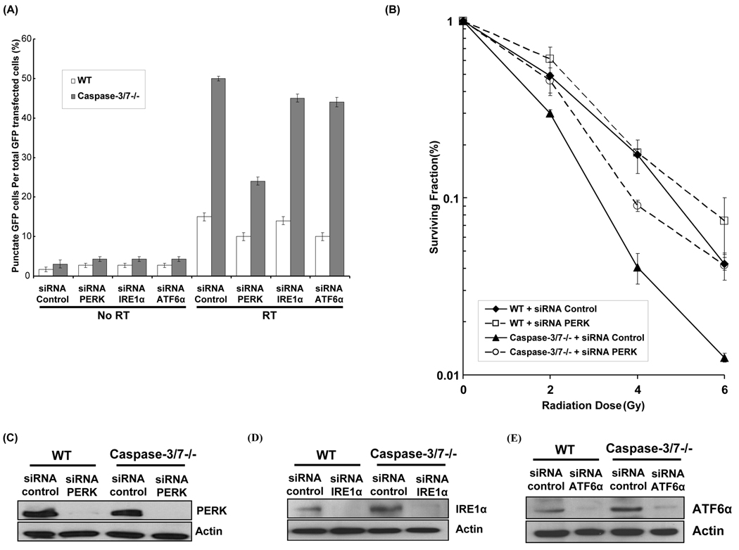
Figure 1. Radiation-induced autophagy is PERK-dependent in caspase-3/7 DKO MEF cells.
(A) Wild-type (WT) and caspase-3/7 DKO MEF cells (caspase-3/7−/−) were transfected with 25nM siRNA against PERK, IRE1α and ATF6α. After 5hrs, cDNA GFP-LC3 (3µg) transfection was performed. The transfected cells were irradiated with 0 Gy or 5 Gy. After 48 hrs the percentage of cells with characteristic punctate GFP-LC3 fluorescence pattern was calculated relative to all GFP-positive cells. This was done in triplicate and error bar is shown as mean ± S.D. (B) Wild-type (WT) and caspase-3/7 DKO MEF cells were transfected with 25nM siRNA against PERK for 24hrs and were irradiated (0–6 Gy). After 8 days, surviving colonies were stained and scored. Values shown are the means ± S.D. of three separate repeated experiments. (C) PERK expression levels were determined by Western blotting using lysates from WT and caspase-3/7 DKO MEF cells treated with siRNA against control and PERK. Actin was probed to demonstrate equal loading. (D) IRE1α expression levels were determined by Western blotting using lysates from WT and caspase-3/7 DKO MEF cells treated with siRNA against control and IRE1α. Actin was probed to demonstrate equal loading. (E) ATF6α expression levels were determined by Western blotting using lysates from WT and caspase-3/7 DKO MEF cells treated with siRNA against control and ATF6α. Actin was probed to demonstrate equal loading.

