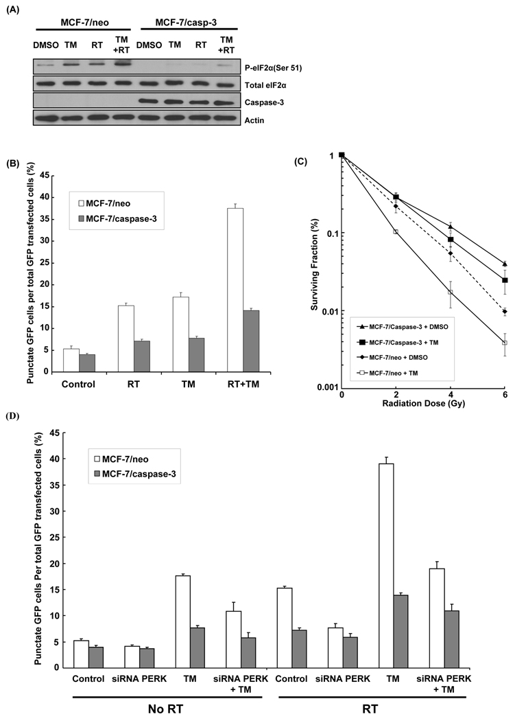
Figure 5. Effects of TM on radiation-induced autophagy and radiosensitivity in a MCF-7 breast cancer cell model.
(A) MCF-7/neo and MCF-7/caspase-3 cells were treated with TM (1 µg/ml), radiation (5 Gy), or combination of both, and were then harvested after 30 min for immunoblotting analyses for P-eIF2α (ser 51), total eIF2α and caspase-3. Actin was probed to demonstrate equal loading. (B) MCF-7/neo and MCF-7/caspase-3 cells were transfected with cDNA GFP-LC3 (3 µg) for 24 hrs and were treated with TM (1 µg/ml for 30 min), radiation (5 Gy) and combination TM (1 µg/ml for 30 min) plus radiation (5 Gy). After 48 hrs, the percentage of cells with punctate GFP-LC3 fluorescence was calculated relative to all GFP-positive cells. This was done in triplicate and error bar is shown as means ± S.D. (C) MCF-7/neo and MCF-7/caspase-3 cells were treated with TM (1 µg/ml) and then irradiated with 0–6 Gy. After 8 days, colonies were stained and scored. Values shown are the means ± S.D. of three separate repeated experiments. (D) MCF-7/neo and MCF-7/caspase-3 cells were transfected with cDNA GFP-LC3 (3 µg) for 24 hrs and were treated with TM (1 µg/ml for 30 min), radiation (5 Gy), siRNA PERK, and combination of these agents. After 48 hrs, the percentage of cells with punctate GFP-LC3 fluorescence was calculated relative to all GFP-positive cells. This was done in triplicate and error bar is shown as means ± S.D.

