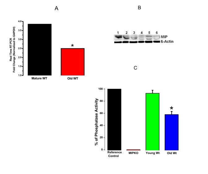
Figure 3. Reduced MIP gene expression, MIP protein levels and MIP phosphatase activity in old skeletal muscles.
(A) Significant reduction in MIP expression in EDL muscles from old Wt mice (red bar) compared with mature Wt mice (black bar). (B) MIP protein content decreased drastically in old skeletal muscle. Lanes 1-3, mature Wt EDL; Lanes 4-6, old Wt EDL; β-actin as controls. (C) MIP enzymatic phosphatase activity reduced by ~ 30% in old Wt EDL muscles as compared to young Wt EDL muscles. * indicates a significant difference (p < 0.01) between the control muscles and a particular genotype.

