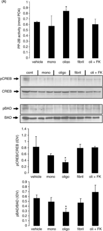
Figure 7. Oligomeric Aβ, but not monomeric or fibrillar Aβ, increases CaN activity and induces CaN-dependent decrease of pCREB and pBAD in rat brain slices.
Rat brain coronal slices (500 mm thick) were maintained in ACSF and treated with monomeric, oligomeric and fibrillar Aβ (0.4 μM) for 3 hrs. A separated set of oligomeric Aβ-treated slices were additionally treated with 10 μM FK506. At the end of the experiment, slices were collected and prepared for measurement of CaN activity (A) and detection of pBAD, BAD, pCREB and CREB by Western blot (B, top). Intensity of specific bands on western blot was analyzed and the result expressed as ratio between phosphorylated protein and the corresponding total protein in the same sample (B, bottom). The experiment was repeated twice with similar results. N=6 in (A) and n=4 (2 for each experiment) in (B). *: p<0.05 vs. vehicle (ANOVA)

