Figure 3.
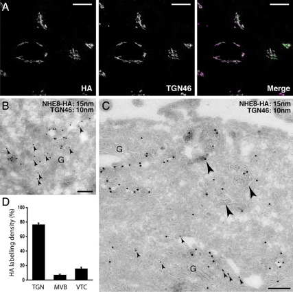
NHE8-HA is localized to the TGN and MVB. (A) HeLa M-cells were stably transfected with NHE8-HA and immunolabeled with anti-HA antibodies or anti-TGN46 antibodies. A series of confocal Z-stacks (Leica optimized) were taken and are displayed as an extended focus view with the merged image in the right panel. Colocalization of TGN46 (magenta) and NHE8-HA (green) appears white in the merged image. Scale bar, 17 μm. (B and C) HeLa M-cells stably expressing NHE8-HA were prepared for immuno-EM and labeled with antibodies to TGN46 (small arrowheads; 10-nm gold) or the HA tag (15-nm gold). (B) TGN46 and NHE8-HA coimmunolabeled vesicles and cisternae of the TGN adjacent to the Golgi stack (G). (C) NHE8-HA immunolabeling could also be identified within MVB (large arrowheads) on ultrathin cryosections. (D) Quantitation of the labeling density of HA associated with the TGN, MVBs, or vesicular/tubular clusters (VTCs) greater than 500 nm from the Golgi stack from three independent immunolabeling experiments ± SEM. Scale bars, 200 nm.

