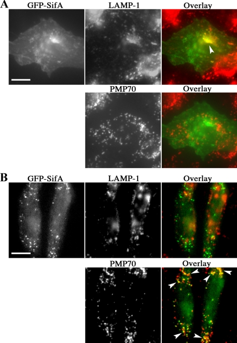
Figure 3.
Distribution of GFP-SifA transfected in mammalian CHO cells. CHO cells were transfected with pEGFP-SifA for 24–48 h., followed by fixation, permeabilization, and immunostaining for the endosome/lysosome marker LAMP-1 and peroxisomal marker PMP70. An overlay of the corresponding images on each row is shown, with yellow color and arrowheads marking areas of overlap with GFP-SifA. Although the majority of cells show the typical codistribution of GFP-SifA and LAMP-1 in A, a small number of cells was found to have GFP-SifA distributed to PMP70 dots (B). Bar, 10 μm.

