Figure 4.
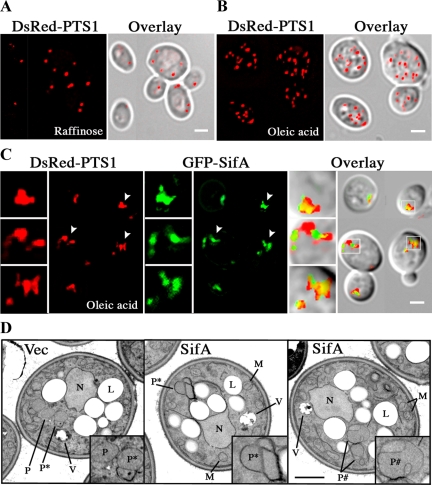
GFP-SifA alters the morphology of peroxisomes. Distribution of peroxisomes (DsRed-PTS1 labeled) in fixed wild-type cells grown in media containing raffinose (A), or oleic acid (B and C). Cells were cotransformed with pDsRed-PTS1 and empty vector (A and B) or pGFP-SifA (C). Overlay indicates merged images of fluorescence and DIC; and yellow indicates the overlap of red and green signals. In C, boxed images to the left of each panel are 2.5× enlargements of smaller areas in each panel indicated by arrowheads or small white squares. Note that peroxisomes associated with SifA are less abundant, tend to clump, and are not always round in shape. The localization of SifA is to one side of peroxisomes and in many cases, appears to bridge nearby clusters of peroxisomes (middle enlarged box). All bars, 2 μm. (D) Cells containing either vector or pGFP-SifA were incubated in oleic acid for 18 h and then fixed and processed for EM. Boxed images at the lower right in each panel show 2× enlargement of specific areas containing peroxisomes that are depicted with letters. P, free and dispersed peroxisomes; P*, clusters of peroxisomes with adherent membranes; P#, enlarged peroxisomes with exaggerated membrane invaginations; N, nucleus; L, lipid body; M, mitochondria; V, vacuole. Bar, 1 μm.

