Figure 1.
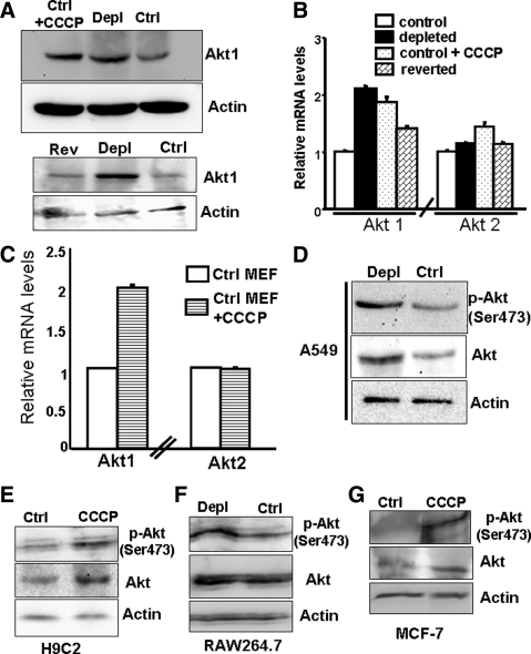
Increased Akt1 protein and mRNA levels in cells subjected to mitochondrial respiratory stress, either by mtDNA depletion or treatment with CCCP. (A) Top, immunoblot analysis of total cell lysate (30 μg protein each) from control, mtDNA-depleted C2C12 cells, and control C2C12 cells treated with CCCP (25 μM, 2 h) using Akt1 antibody (1:1000 dilution). Bottom, immunoblot analysis of total cell lysates (30 μg each) from control, mtDNA-depleted, and reverted C2C12 cells using Akt1 antibody. β-Actin was used as an internal loading control. (B) Real-time PCR analysis of mouse Akt1 and Akt2 mRNA levels in control, mtDNA-depleted, and reverted C2C12 cells and also in control C2C12 cells treated with CCCP (25 μM, 2 h). (C) Real-time PCR analysis of Akt1 and Akt2 mRNA in mouse embryonic fibroblasts (MEFs) treated without and with CCCP (25 μM, 2 h). (D) Immunoblot analysis of total cell extracts (30 μg each) of control and mtDNA-depleted A549 lung carcinoma cells with antibody to Akt and phospho-Ser473 Akt. (E–G) Immunoblot analysis of nuclear extracts from H9C2 cells treated with CCCP, mtDNA-depleted RAW 264.7 cells, and CCCP treated MCF-7 cells with Akt and Ser437 pAkt antibodies. Although not shown, mtDNA contents of A549 and MCF-7 cells were ∼20–25% of control cells. In B and C, mean ± SEM values were derived from three to four separate experiments.

