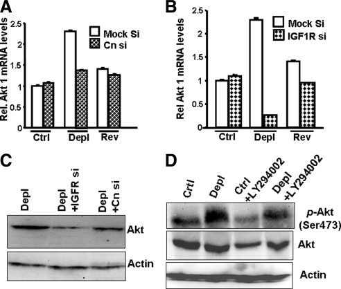
Figure 3.
The role of calcineurin, IGF1R and PI3-K in the activation of Akt1 in cells subjected to mitochondrial stress. (A) Effects of CnAα mRNA knockdown on the levels of Akt1 mRNA in control, mtDNA-depleted, and reverted C2C12 cells. (B) Effects of IGF1R mRNA knockdown on the levels of Akt1 mRNA in control, mtDNA-depleted, and reverted C 2C12 cells. In both A and B, Akt1 mRNA levels were quantified using real-time PCR analysis. Mean ± SEM values were calculated from three separate estimates. (C) Immunoblot analysis of total cell lysates (50 μg each) from control, mtDNA-depleted C2C12 cells, and control as well as depleted cells expressing siRNAs to CnAα and IGF1R were developed with Akt antibody. A companion blot was probed with actin antibody to assess loading levels. (D) Effects of the PI3-K inhibitor LY294002 (50 μM, 2 h) on Akt and phospho-Ser473 Akt in control and mtDNA-depleted cells. The immunoblot represents the analysis of total lysates (50 μg protein each) developed with the indicated antibodies. A companion blot was probed with actin antibody to assess protein loading.

