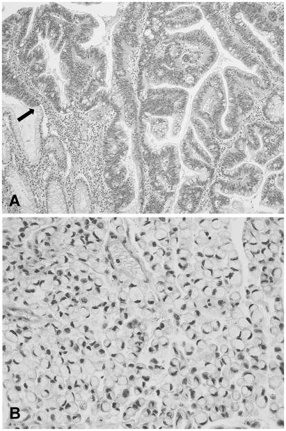
Figure 3.
Microphotographs of gastric adenocarcinoma. A) Intestinal type, showing tumor cells cohesively arranged forming irregular glandular structures. On the left lower corner there are few glands with intestinal metaplasia. An arrow shows the transition zone between intestinal metaplasia and adenocarcinoma (×100). B) Diffuse type, with tumor cells that show lack of cohesiveness infiltrating diffusely. In this subtype, the signet-ring adenocarcinoma, the nuclei are pushed to the periphery due to the abundant mucinous cytoplasmic content (×400).

