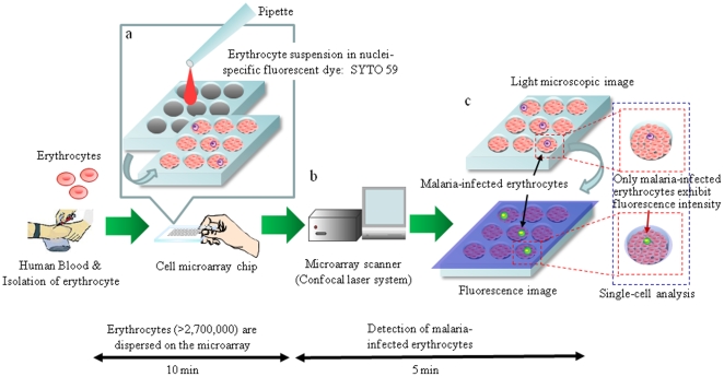
Figure 1. Schematic process for detection of malaria-infected erythrocytes on a cell microarray chip.
(a) Erythrocytes stained with a nuclei-specific fluorescent dye, SYTO 59, for the staining of malaria nuclei were dispersed on a cell microarray chip using a pipette, which led to the formation of a monolayer of erythrocytes in the microchambers. (b) Malaria-infected erythrocytes were detected using a microarray scanner with a confocal fluorescence laser by monitoring fluorescence-positive erythrocytes. (c) The target malaria-infected erythrocytes were analyzed quantitatively at the single-cell level.

