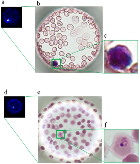
Figure 5. Discrimination of leukocytes and malaria-infected erythrocytes on a cell microarray chip.
(a) Scanned image of leukocytes on a microarray chip obtained with a microarray scanner. (b) Leukocytes were identified by Giemsa staining. (c) Magnified view of the boxed region. (d) Microarray scanning image of malaria-infected erythrocytes. (e) Malaria-infected erythrocytes were confirmed by Giemsa staining after microarray scanning. (f) Magnified view of the boxed region.

