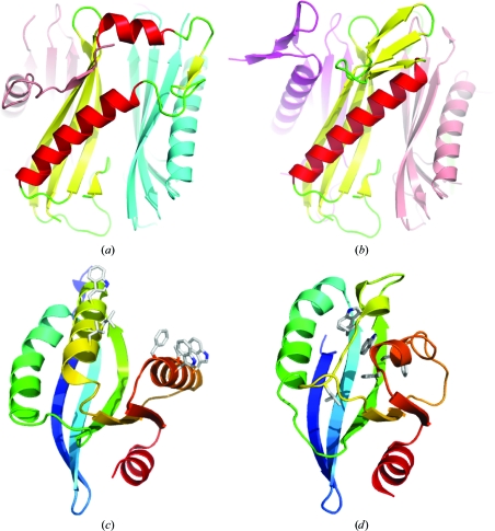
Figure 7.
Metamorphic proteins. (a, b)Side-by-side comparison of alternatively folded subunits of the DUF74 pentamer (PDB entry 1vr4). Chain A and chain D (b) are coloured according to their secondary structure. The adjacent subunits are coloured as follows: chain B, light blue; chain C, magenta; chain E, pink. (c, d) Side-by-side comparison of the Sfri0576-like family structures 2q3l (c) and 2ook (d). The equivalent nonpolar residues that are exposed on the 2q3l surface and buried in the 2ook core are shown in stick representation.

