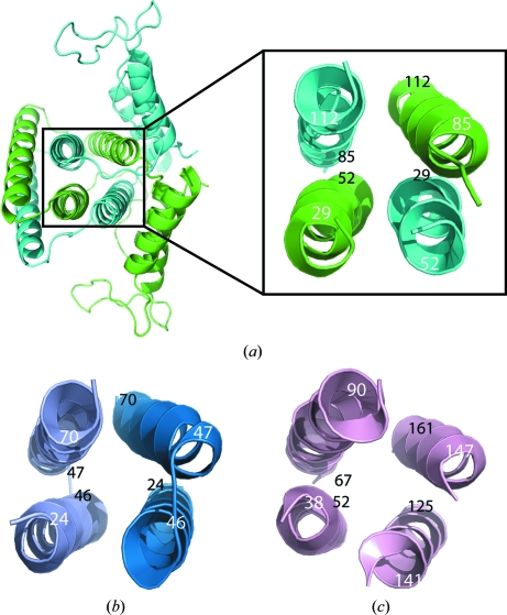
Figure 4.
Comparison of the core four-helix bundles from the α-helical NTPase superfamily. These four-helix bundles either assemble upon dimerization or are present in a single monomer, resulting in the same down–up–down–up topology. White numbers are closest to the viewer and black numbers are farthest away. (a) Ribbon diagram showing the dimer of YP_001813558.1. (b) Ribbon diagram showing the central four helices of S. solfataricus MazG. (c) Ribbon diagram showing the central four helices from a single protomer of C. jejuni dUTPase.

